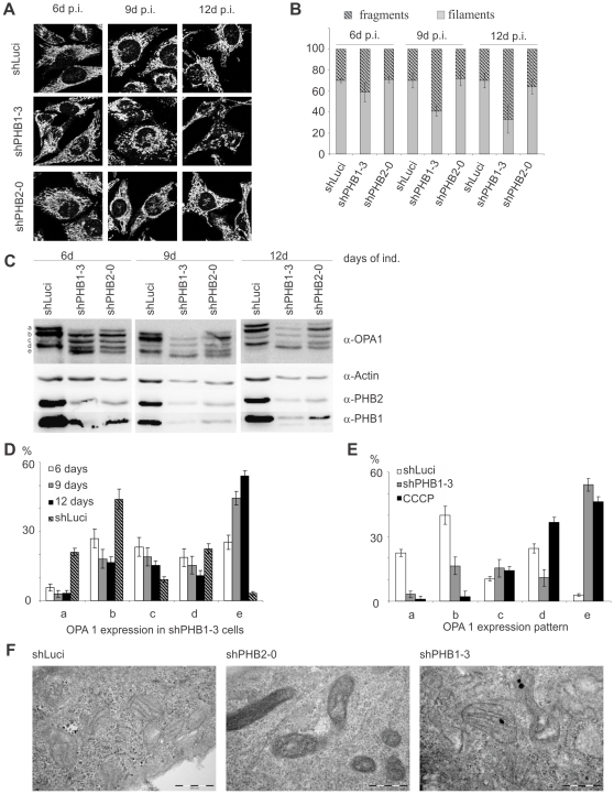
Figure 3. Prohibitin knockdown led to fragmentation of the mitochondrial network without affecting cristae morphology.
(A, B) Mitochondria from prohibitin knockdown cells were stained with anti-Tom 20 and mitochondrial fragmentation was quantified using ImageJ software. An extended induction with doxycycline led to an increased fragmentation in shPHB1-3 expressing cells. (C) Western blots showing that prolonged doxycycline-induced shPHB1-3 expression led to a stronger prohibitin knockdown than shPHB2-0 expression, correlating with an increasing fragmentation of the fusion competent fragments a and b of OPA1. (D) Quantification showed that the fusion competent forms were degraded into the smaller fragments c and e. (E) Quantification of OPA1 patterning in control cells (shLuci), PHB1-depleted cells (shPHB1-3) and cells treated with 60 µM CCCP for 80 min (CCCP) to induce loss of ΔΨm. The OPA1 pattern in CCCP-treated cells differed from shPHB1-3 expressing cells, as the fragments a and b were completely lost while the smaller fragments d and e were increased. (F) TEM pictures of cells with a prohibitin knockdown induced for 10 days showed a more dense appearance of mitochondria than shLuci-expressing control cells, but the morphology of the cristae structure was not changed.

