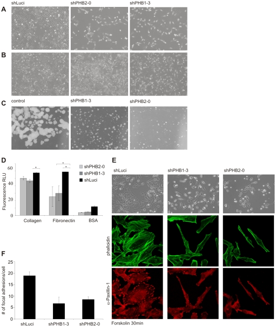
Figure 4. Prohibitin knockdown led to a changed morphology in HeLa cells and to reduced cell-cell contact.
(A, B) Silencing prohibitin expression for 10 days resulted in a stretched cell morphology with the nucleus slightly raised. (A) When seeded at low density prohibitin knockdown cells remained as single cells with minimal cell-cell contact. (B) At higher seed density, prohibitin-knockdown cells still showed an elongated morphology but either formed clusters (shPHB1-3) or remained predominantly as single cells with reduced cell-cell contact. The proliferation rate of prohibitin knockdown cells was also increased in comparison to cells seeded at low to normal density. (C) Preventing anchorage by seeding cells on agar-coated tissue culture plates strongly reduced proliferation rate and colony formation in prohibitin knockdown cells, whereas control cells (shLuci expressing cells) formed large cell clumps. (D) Prohibitin-depleted and control cells were treated with CFSE to increase fluorescence intensity and seeded on extracellular matrix proteins collagen or fibronectin, or on BSA for 30 min. Non- adherent cells were washed off and fluorescence intensity was measured. Prohibitin knockdown significantly reduced adhesion. (E) Doxycycline-induced cells were seeded on coverslips for 20 h followed by serum starvation for 4 h. After treatment with 50 µM Forskolin for 20 min, brightfield images showed lamellipodia formation in control cells but not in prohibitin- depleted cells. Furthermore, prohibitin-depleted cells showed decreased formation of focal adhesions as shown by immunocytochemistry staining using phalloidin and anti-paxillin antibodies. (F) For the quantification of focal adhesions in control and prohibitin knockdown cells, α-paxillin 1 stained spots were counted in 30 cells per condition.

