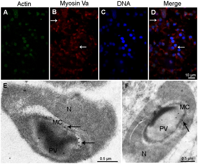
Figure 5. Localization of myosin Va at the mid stage during E. sinensis spermiogenesis.
(A–D) Immunofluorescent localization of myosin Va in early mid stage. Myosin Va staining can be seen at the proacrosomal vesicle (PV) (arrows in B, D). Scale bar = 10 µm. (E–F) Immunogold labeling of myosin Va is present in the membrane complex (MC) and the proacrosomal vesicle (PV) membrane at late mid stage (arrows in E, F). E shows longitudinal section and F shows cross section of mid stage spermatid.

