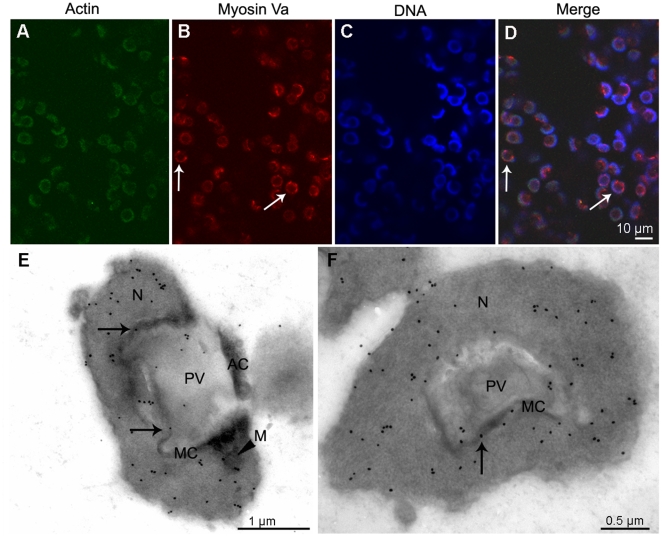
Figure 6. Localization of myosin Va at the beginning of late stage during E. sinensis spermiogenesis.
(A–D) Immunofluorescent localization of myosin Va. Myosin Va concentrates in the nucleus (arrows in B and D). (E–F) Immunogold labeling of myosin Va. Prominent myosin Va labeling is present in the nucleus (N), and some associates with the nuclear membrane and the membrane complex (MC) (arrows in E, F), as well as mitochondria (M) (arrowhead in E). E. longitudinal section and F. cross section of the initiation of late stage.

