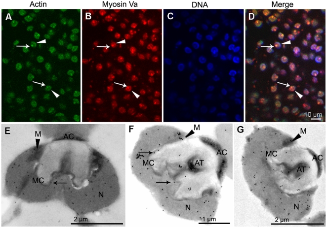
Figure 7. Localization of myosin Va at the late stage during E. sinensis spermiogenesis.
(A–D) Immunofluorescent localization of myosin Va. Myosin Va distributes in the nucleus (N) (arrowhead in B,D) and acrosomal tubule (AT) (arrow in B,D), actin staining is present in the nucleus (N) (arrowhead in A,D) and acrosomal tubule (AT) (arrow in A,D), myosin Va colocalizes with actin (D). (E–G) Immunoelectron microscopy analyses of myosin Va. (E) When the acrosomal tubule (AT) begin to form, myosin Va associates with the nuclear membrane and the membrane complex (MC) (arrow in E). (F) After the acrosomal tubule (AT) formation, myosin Va bounds to the nuclear membrane and localizes in the membrane complex (MC) (arrows in F) and acrosomal tubule (AT). (G) Myosin Va localization in mature spermatozoon. Myosin Va distributes in the nucleus (N) and acrosomal tubule (AT). Mitochondria (M) are decorated with myosin Va labeling (arrowheads in E–G).

