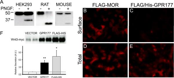
Figure 5. GPR177 structure and function.
(A) Crude microsomal membrane preparations from HEK 293 cells transfected with FLAG/6× His-tagged GPR177 cDNA (HEK293), rat brain (RAT), and mouse brain (MOUSE) were digested in the presence (+ lanes) or absence (− lanes) of N-glycosidase F (PNGF). Lysates were resolved by SDS-PAGE and probed with anti-GPR177 antibodies. Data are representative of three separate experiments. (B–E) HEK 293 cells transfected with FLAG-tagged MOR (FLAG-MOR) or N-terminal FLAG/6× His tagged-GPR177 (FLAG/His-GPR177) cDNA and labeled with anti-FLAG antibodies under permeabilized (Total) and non-permeabilized (Surface) conditions. Representative images from two independent experiments are shown. (F) HEK 293 cells were cotransfected with myc-tagged Wnt3 cDNA in pCMV-Tag3B vector and either pCMV-Tag2B vector (VECTOR), untagged GPR177 cDNA in pCMV-Sport6 vector (GPR177), or FLAG/6× His-tagged GPR177 in pCMV-Tag2B vector (FLAG-HIS). Wnt3-myc secretion was assayed as described in Experimental Procedures. Western blots were probed with anti-myc antibodies, and bands were quantitated using ImageJ software (NIH). Data was subjected to two-tailed Student's t-test. Dotted line indicates secretion obtained with empty vector control and assigned a relative value of 1.0 (n= 4–6, **p<0.01, *p<0.05).

