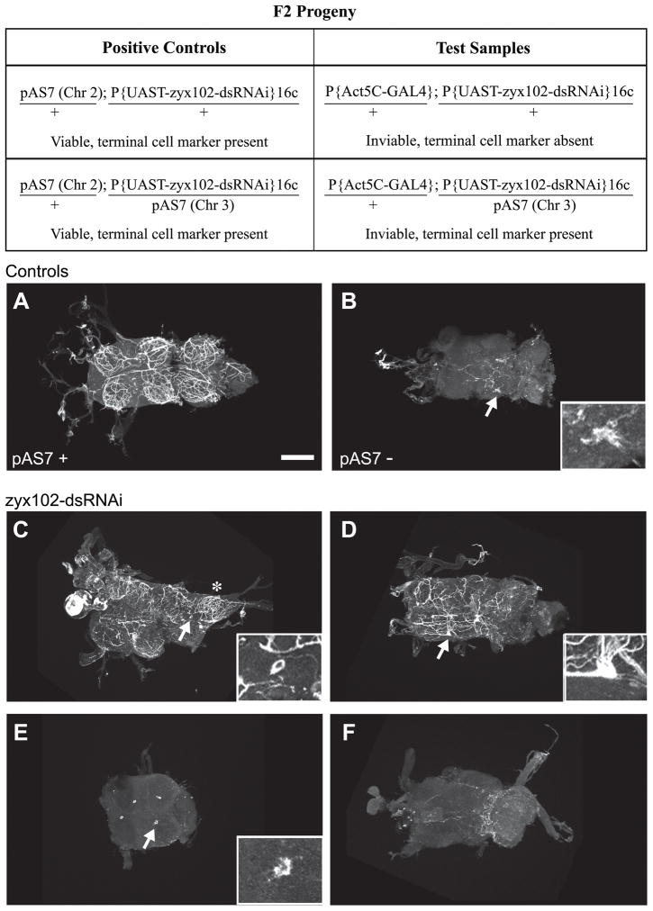
Figure 8. Tracheal terminal cells elaborate processes in the Zyx102-knockdown flies.
To investigate whether tracheal cells in the VNC are present if RNA-interference is used to reduce the levels of Zyx102, and to see to what extent these tracheal cells develop processes that undergo normal air-filling, a GFP-tagged marker for differentiated terminal cells of the tracheal system (pAS7) was used. VNCs were prepared from pharate or freshly eclosed adults and processed for immunostaining using anti-GFP. Top panel: F2 progeny from a single cross were used as test samples and positive controls. Genotypes for chromosomes 2 and 3 are shown; viability and presence or absence of the pAS7 marker are indicated. Half of the inviable pharate adult flies should be negative for GFP, half positive for GFP. These flies cannot a priori be distinguished from each other. The P{UAS-zyx102-dsRNA}16c stock was used as a negative control. Refer to Table 1 for further description of experimental observations. Arrows indicate areas shown in insets. Anterior is to the left. Bar = 100 μm.
A. Positive control: a VNC dissected from a viable pharate adult carrying the tracheal system terminal cell marker, pAS7 and P{UAS-zyx102-dsRNA}16c. The fine network of processes tagged with the terminal cell marker is nearly identical to the tracheal network observable because of air-filling (Fig. 7).
B. Negative control: a VNC dissected from a viable pharate adult carrying P{UAS-zyx102-dsRNA}16c only. Note the background evident as either fine processes or a larger aggregate (arrow and inset). Although two out of five samples showed no labeling, three out of five showed patterns like this one.
C–F. VNCs dissected from inviable pharate adults (thus carrying both Act5C-GAL4 and UAS-zyx102-dsRNA; half are expected to carry the terminal cell marker, pAS7, half should not). Some lobes exhibit a normal tracheolar network (asterisk in C), while others do not. In C and D, terminal cell bodies can be detected (arrow and inset), some with (D) and some without (C) labeled processes. In some VNCs dissected from inviable adults, single cells were strongly labeled with the terminal cell marker (arrows and insets, E), a pattern not seen in negative controls (data not shown). The VNC in E lost two of the six lobes during the staining procedure. The labeling pattern in F is strongly akin to that seen in the negative controls, and is likely from a specimen not marked with pAS7.

