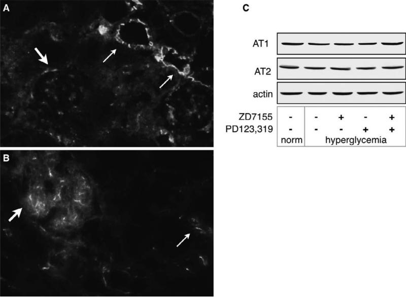
Figure 1. Angiotensin receptors are expressed in normal murine kidneys.
AT1 (A) and AT2 (B) were detected in kidney slices as described in the methods section. Arrows indicate localization of AT1 and AT2. Magnification = 20X. AT1 is strongly expressed in the apical and lateral membranes of the tubules (thin arrows), and in mesangial cells (thick arrow),.AT2 is expressed mostly in glomerular endothelial cells (thick arrow) and in the proximal tubules (thin arrow). The pictures are representative of kidneys from 2 individual mice. C) detection of AT1 and AT2 by western blot in kidney cortex homogenates using the same antibodies as in A and B.

