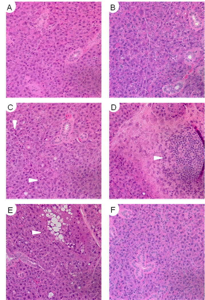
Figure 4. Histological analysis of parotid salivary gland structure 90 days after fractionated radiation.
Parotid salivary glands from mice treated in figure 3 were paraffin embedded and sections were stained with hematoxylin and eosin. A) Untreated FVB, B) Untreated myr-Akt1, C) Irradiated FVB, D) Irradiated FVB, E) Irradiated myr-Akt1, F) Irradiated FVB with IGF-1 injections. Images A, B, C, and F are representative of the overall tissue architecture.

