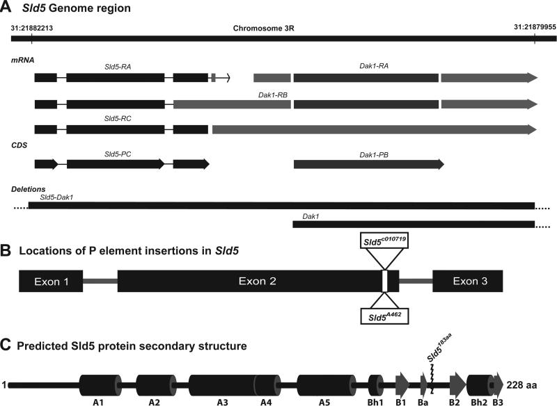
Figure 1. Sld5 Gene Region, P-element insertion, and Protein layout.
A. Sld5 gene is located on chromosome 3R and may be part of a multicistronic mRNA that also contains Dak1. Two different deletion lines were used to show that Sld5 mutations were specific to Sld5 and not also defective for Dak1. B. Map of the Sld5 gene showing insertion site of the two independent P element lines used in this study. C. Map of the predicted 1° protein structure of Sld5 based on alignment with the human protein [9,22,23] comprised of a N terminal “A” domain comprised mainly of alpha helices and a C terminal “B” domain with 4 beta sheet regions. The insertion of the respective P elements truncates the protein removing critical residues in the B domain required for stability of the GINS complex [12].

