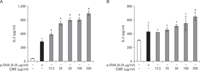
Figure 1.
Effects of CME on the cross-presentation of exogenous OVA in DCs. (A) DC2.4 cells and (B) BM-DCs were incubated with the indicated amounts of CME for 2 hrs, and then combined with OVA-microspheres. After 6 hrs incubation, the cells were washed, fixed, and the amounts of OVA peptides presented on MHC class I molecules were assessed using OVA-specific CD8 T cell hybridoma, CD8OVA1.3. The amounts of IL-2 produced from OVA-specific CD8 T cells were assayed by a commercial IL-2 ELISA kit. Data have been presented as means±S.D. of three independent experiments. *p<0.05 vs. cells only; †p<0.05, ‡p<0.01 vs. OVA only.

