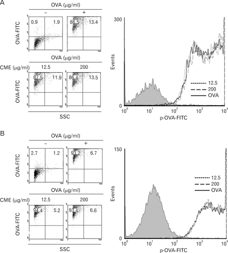
Figure 3.
Effects of CME on the phagocytic activity. (A) DC2.4 cells and (B) BM-DCs were cultured with CME for 2 hrs, and then combined with microspheres containing both OVA and FITC (6.25µg/ml). After 2 hrs incubation, unphagocytozed microspherses were washed, and the cells were harvested by gentle pipetting, and then analyzed by flow cytometry.

