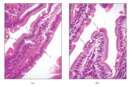
Figure 2.
Histological duodenum sections of rats fed standard (based on egg white) diet (a) and CryIa12 toxin-containing diet (b) (600x amplification). Arrow 1 shows that the edge formed by microvilli remains intact for all treatments. Arrow 2 shows the nucleus of enterocytes, normal in all treatments. Arrow 3 shows the goblet cells with normal size for all treatments. The tissues were stained with hematoxylin-eosin.

