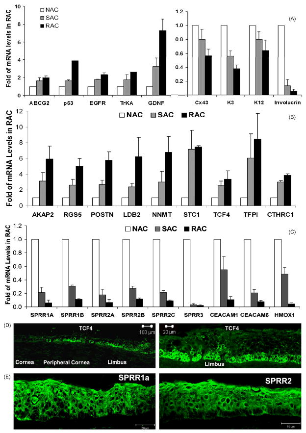
Figure 3.
Validation of 27 genes for stem cell phenotype. The expression patterns of known 5 stem cell associated and 4 differentiation markers (A), 9 highly up- (B) and 9 highly down- (C) regulated new genes were verified by RT and quantitative real-time PCR in the isolated RAC, SAC and NAC populations from limbal epithelial tissues obtained from separate adhesion experiments. The representative images of immunofluorescent staining on corneal limbal tissue frozen sections showing immunolocalozation of TCF4 that was positive only at basal cells of limbal and peripheral corneal epithelia (D) or SPRR1a and SPRR2 that were negative at limbal basal cells (E).

