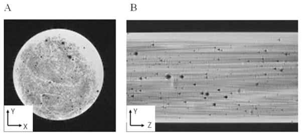
Figure 4.
High-resolution MR images of the fabricated tissue surrogate (20% glass-fiber volume in a 1% agarose hydrogel) clearly showing the aligned glass-fiber structure. The transverse image (left) had a 5 × 5 mm2 field of view, whereas the axial image (right) had a 5 × 7.6 mm2 field of view (truncated for display). The black dots correspond to microscopic air bubbles. The voxel resolution was 40 × 40 × 40 μm3.

