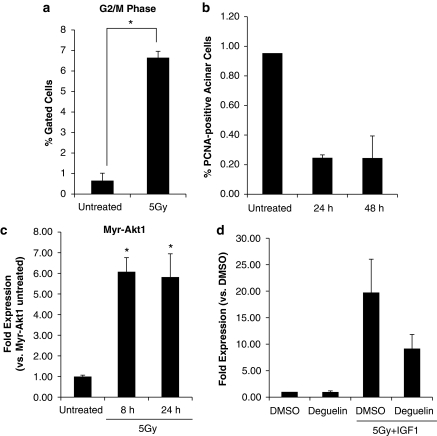
Figure 2.
Radiation induces cell cycle arrest in myr-Akt1 mice. The head and neck regions of myr-Akt1 mice were irradiated (a–c). Parotid glands were removed 8, 24 and 48 h after treatment. (a) In all, 8 h tissues were dispersed, stained with propidium iodide and analyzed by flow cytometry. The data are shown as the mean percentage of gated cells in G2/M+S.E.M. of ⩾3 mice per treatment. (b) In total, 24 and 48 h tissues were embedded in paraffin and stained for PCNA. The graphs represent the number of PCNA-positive acinar cells as a percentage of total acinar cells counted. The data are shown as the mean+S.E.M. (c) RNA was isolated from 8 and 24 h tissues, and real-time RT-PCR was run with primers to amplify p21. Results were calculated using the 2−ΔΔCt method, normalized to wild-type untreated and shown as the mean+S.E.M. of ⩾3 mice per treatment. *A significant difference (P⩽0.05) between untreated and irradiated glands. (d) Wild-type mice were treated intraperitoneally with deguelin (4 mg/kg) immediately before IGF1 injections and head and neck irradiation. Parotid glands were removed after 8 h, RNA was isolated, and real-time RT-PCR was run with primers to amplify p21. Results were calculated using the 2−ΔΔCt method, normalized to vehicle control (DMSO alone) and shown as the mean+S.D.

