Figure 5.
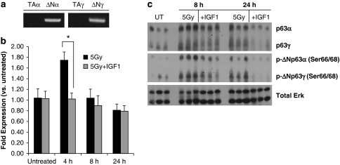
ΔNp63 mRNA and protein expression increases in irradiated parotid glands. (a) RNA was isolated from untreated parotid glands, and RT-PCR was run with primers to amplify p63 isoforms (TAα, ΔNα, TAγ and ΔNγ). PCR products were electrophoresed on a 1% agarose gel. (b) The head and neck regions of wild-type mice were irradiated ±IGF1 pretreatment. Parotid glands were removed, RNA was isolated and real-time RT-PCR was run with primers to amplify total p63. Results were calculated using the 2−ΔΔCt method, normalized to untreated and shown as the mean+S.E.M. of ⩾3 mice per treatment. *A significant difference (P⩽0.05) between irradiated glands ±IGF1 pretreatment as measured by a Student's t-test. (c) The head and neck regions of wild-type mice were irradiated ±IGF1 pretreatment. Parotid glands were removed, protein lysates were prepared and western blotting was performed as described in Materials and methods. Total ERK was used to confirm equal loading of lanes

