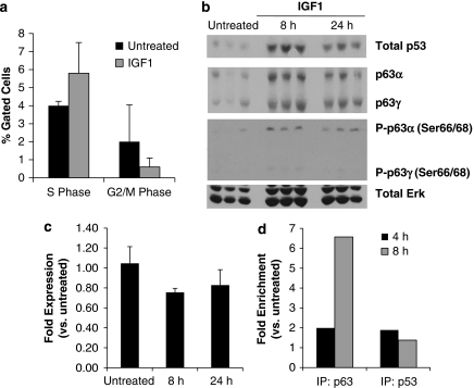
Figure 8.
IGF1-induced effects after irradiation are not detected in unstressed salivary glands. Wild-type mice were treated with IGF1, and parotid glands were removed after 4, 8, and 24 h. (a) Cell cycle distribution was analyzed for 8 h tissues as described for Figure 1a. The data are shown as the mean percentage of gated cells in G2/M phase+S.E.M. of ⩾3 mice per treatment. (b) In all, 8 and 24 h protein lysates were analyzed as described for Figure 3. (c) p21 mRNA expression was measured after 8 and 24 h as described for Figure 4. (d) ChIP was performed on 4 and 8 h tissues with an antibody recognizing total p63 or total p53. Precipitated DNA was analyzed and graphed as described for Figure 6. Normalized values are shown as fold versus untreated and represent trends that were consistent between multiple independent experiments

