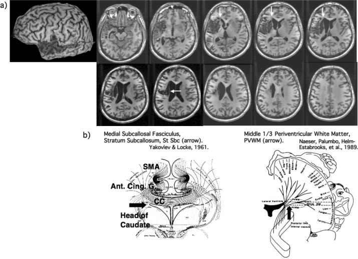
Figure 1.
a) Structural MRI scan (3-dimensional magnetization prepared rapid gradient echo) obtained at 1 year poststroke. Large cortical lesion was present in the left temporal lobe, including anterior portions of superior and middle temporal gyrus, with posterior extension across most of the middle temporal gyrus (BA 21). Only small lesion was present in the more anterior portion of Wernicke's area. Small cortical lesion was present in only the most inferior portions of Broca's area. The lesion was primarily subcortical, with lesion in the two, deep white matter areas near ventricle, associated with persistent nonfluent speech: 1) medial subcallosal fasciculus area, deep to Broca's area and adjacent to frontal horn (vertical white arrows); and 2) periventricular white matter area adjacent to body of lateral ventricle, deep to sensori-motor mouth area (horizontal white arrow). b) Diagrams showing location, and some pathways within each of these two white matter areas adjacent to ventricle: 1) medial subcallosal fasciculus area adjacent to frontal horn, showing pathways from SMA and anterior cingulate gyrus to head of caudate (horizontal black arrow); and 2) periventricular white matter (PVWM) adjacent to body of lateral ventricle (vertical black arrow). See also text, where additional pathways are listed.

