Figure 3.
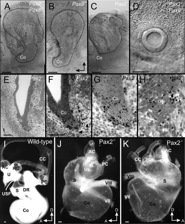
Overview of ear defects and sections. Whole mounted (A-D, I-K) and coronally sectioned ears (E-H) show the mutant effects on morphology. The cochlear duct is truncated in Pax2 null whereas Pax2;8 double null mice have only an otic vesicle at E11.5 (A, B; outlined by a dotted line). Coronal sections (see vertical line in C) show no cochlear outgrowth in Pax2 null mice as late as E13.5 (E). Numerous apoptotic cells in and around the cochlear duct (F, G) are indicated by chromatin condensation. Cartilage is obvious lateral but not medial to the ear (E). Higher magnification shows also apoptotic cells in the ganglion including in the more ventral, presumably spiral ganglion part (H). At E18.5 wild-type ears have developed a coiled cochlea, three distinct canals and recesses for the saccule and the utricle (I). The utricle is separated by the utriculo-saccular foramen from the saccule (USF in I) and the cochlea is separated from the saccule by the ductus reunions (DR in I). Pax2 null mice have an inflated saccule and cochlear duct with shortened semicircular canals (J, K). The only constriction resulting in two adjacent vesicles is near the saccule. Facial (VII) and vestibular (VIII) nerves are abutting the ear adjacent to the constriction separating the superior from the inferior division. AC, anterior canal/crista; CC, common crus; HC, horizontal canal/crista; Co, cochlear duct/sack; DR, ductus reunions; Ggl, ganglion; PC, posterior canal/crista; S, Saccule; U, utricle; USF, utriculo-saccular foramen; VII, facial nerve; VIII vestibular/cochlear nerve. Bar indicates 100 um in A-F, I-K, 10 um in G, L.

