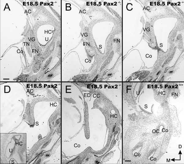
Figure 4.
Overview of sections through E18.5 ear. These near serial coronal sections through the ear show the unusual morphology with a vesicle within the otic capsule (top of each section) and the large ventral vesicle that is expanded into the brain cavity, labeled as cochlea (C in A-E) instead of the multiple cross sections through the cochlear duct found in wild-type (F). Note the brain has been removed to verify before sectioning that a vesicle was present. Canals and canal cristae can be found along the superior vesicle surrounded by periodic mesenchyme. However, the horizontal crista is not in the horizontal canal (HC in A, D) whereas it sits in wild-type at the orifice of the horizontal canal (insert in D). Hair cells of the utircular and saccular macula are continuous and extend into the ventral, extruded sack of the ear (A-D). While adjacent to each other, the endolymphatic duct (ED) and the common crus (CC) are nevertheless distinct anatomical entities (E). AC, anterior canal/crista; CC, common crus; FN, facial nerve; ED, endolymphatic duct; HC, horizontal canal/crista; Co, cochlea duct/sack; OC, organ of Corti; PC, posterior canal/crista; S, saccule; TN, trigeminal nerve; U, utricle; VG, vestibular ganglion. Bar indicates 100 um.

