Figure 7.
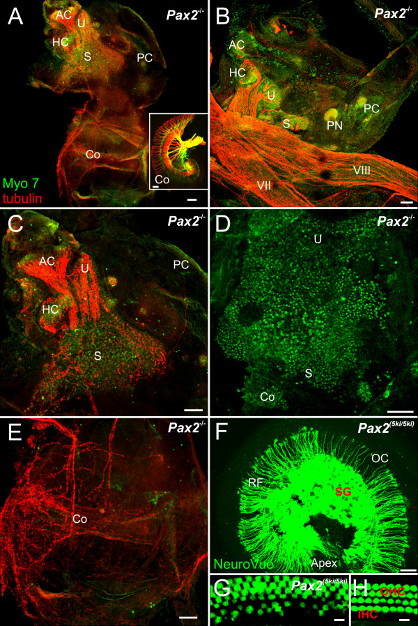
Vestibular hair cells and some innervation develop in Pax2 null mice. Pattern of innervations and distribution of hair cells as revealed by anti-tubulin (red) and anti-Myo 7 (green) antibodies (A-E, G, H) and lipophilic dye tracing (insert in A, F). Hair cells are concentrated to the superior part of the ear in Pax2 null mice (A, C, D), innervations exists to vestibular (A-C) and cochlear sack (A, E). Pax2 5ki/5ki show a near normal innervation of the cochlea (F) compared to that found in wild-type (insert in A). Anterior, horizontal and posterior canal and crista can be recognized in all ears (compare C, F) and may receive no or few nerve fibers. Myosin 7 labeled hair cells form an unstructured aggregation tentatively divided into utricle (U) saccule (S). In addition to several small isolated patches of hair cells we found aeas without hair cells near the ventral border of the saccular patch (A-D). Innervation implies that nerve fiber pathfinding is unaltered. The cochlear sack has no hair cells and a diffuse innervation. This contrasts to the radial fibers (RF) to the inner and outer hair cells (IHC, OHC) of the organ of Corti (OC) with the in Pax2 5ki/5ki (F, G) and wild-type cochleae (insert in A, H). AC, anterior canal crista; HC, horizontal canal crista; IHC, inner hair cell; OC, organ of Corti; OHC, outer hair cell; PC, posterior canal crista; RF, radial fibers; S, saccule; SG, spiral ganglion; U, utricle. Bar indicates 100 um.

