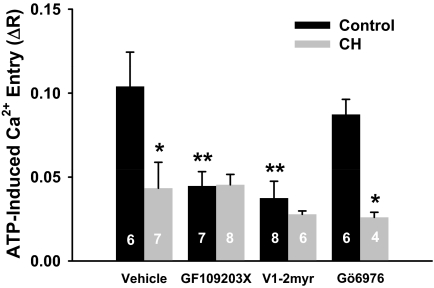
Fig. 3.
PKC inhibition blunts ATP-induced Ca2+ influx in endothelium from controls but not from CH pulmonary arteries. ATP-induced Ca2+ entry (ΔR) was assessed after the SOC response in the presence of vehicle, the nonselective PKC inhibitor GF109203X (1 μM), the PKCε inhibitor V1-2myr (10 μM), or the PKCα/β inhibitor Gö6976 (6 nM). Values are mean ± S.E. n is the number of endothelial sheets (40–100 cells/sheet) and is indicated within the data bars. P ≤ 0.05: *, versus control vehicle; **, versus control vehicle.

