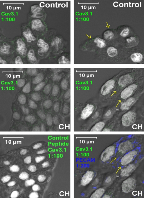
Fig. 7.
Immunofluorescence of Cav3.1 (α1G) T-type VGCC subunit (green) in freshly isolated endothelial cells from small pulmonary arteries harvested from control (top) and CH (middle and bottom) rats (magnification: ×630). Top and middle, images for control (top) and CH (middle) show detectable Cav3.1 channel subunit fluorescence (indicated by yellow arrows). Bottom left, coincubation with the blocking peptide prevented Cav3.1 immunofluorescence. Bottom right, positive PECAM-1 immunofluorescence is shown in blue. Cav3.1 and Sytox nuclear stain is in white in all cases.

