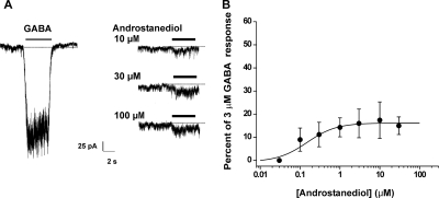
Fig. 5.
Direct activation of GABAA receptor-mediated inward currents by androstanediol in acutely dissociated CA1 pyramidal neurons. A, representative traces comparing responses of GABA (3 μM) and androstanediol (10–100 μM) in the same cell. The interval between the GABA and androstanediol perfusions was 30 s. B, peak direct androstanediol (0.1–100 μM)-induced current expressed as a fraction of the mean current amplitude evoked by 3 μM GABA. There was modest (<25%) direct activation even at highest concentrations (100 μM). The curve show arbitrary logistic fits to the mean values; parameters could not be derived because responses were largely saturated at concentrations <10 μM, and this is probably due to slow onset and duration of the currents. Each point represents the mean ± S.E.M. of data from four to six cells.

