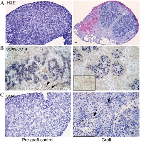
Figure 1.
(A) Haematoxylin and eosin. Seminiferous cord formation in first-trimester human fetal testis xenografts. Pre-graft control testes (left) lacked completed seminiferous cord formation, while grafts had developed seminiferous cords with normal appearance (right). (B) Identification of cell types within newly formed seminiferous cords in first-trimester human fetal testis xenografts (right) in comparison with pre-graft controls (left) based on immunoexpression of SOX9/OCT4. Isolated OCT4+ (brown) germ cells can be identified (arrowheads) in areas devoid of SOX9 (blue) expressing Sertoli cells in pre-graft controls, but after grafting all germ cells are enclosed within clearly defined seminiferous cords. (C) The cords are separated from the interstitial compartment by a basement membrane outlined by the expression of SMA, (arrows), while the pre-graft control tissue is negative for SMA. Scale bar = 50 µm. Negative controls are also shown (inset).

