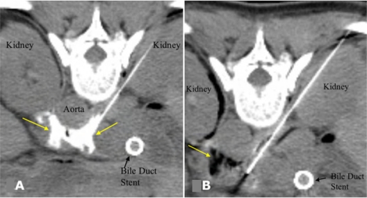
Figure 3.
A) CT image after injection of a small volume of dilute contrast agent through both needles, confirming correct distribution of injected contrast around the celiac axis (arrows) prior to alcohol injection. B) After injection of alcohol, darkened region (arrow) shows its distribution in the vicinity of the celiac plexus. Copyright © 2007. Reproduced with permission from Arellano RS. Image-guided pain management, Part 1: celiac plexus block for palliative pain relief. Radiology Rounds, Vol 5. Boston, MA: Massachusetts General Hospital; 2007.

