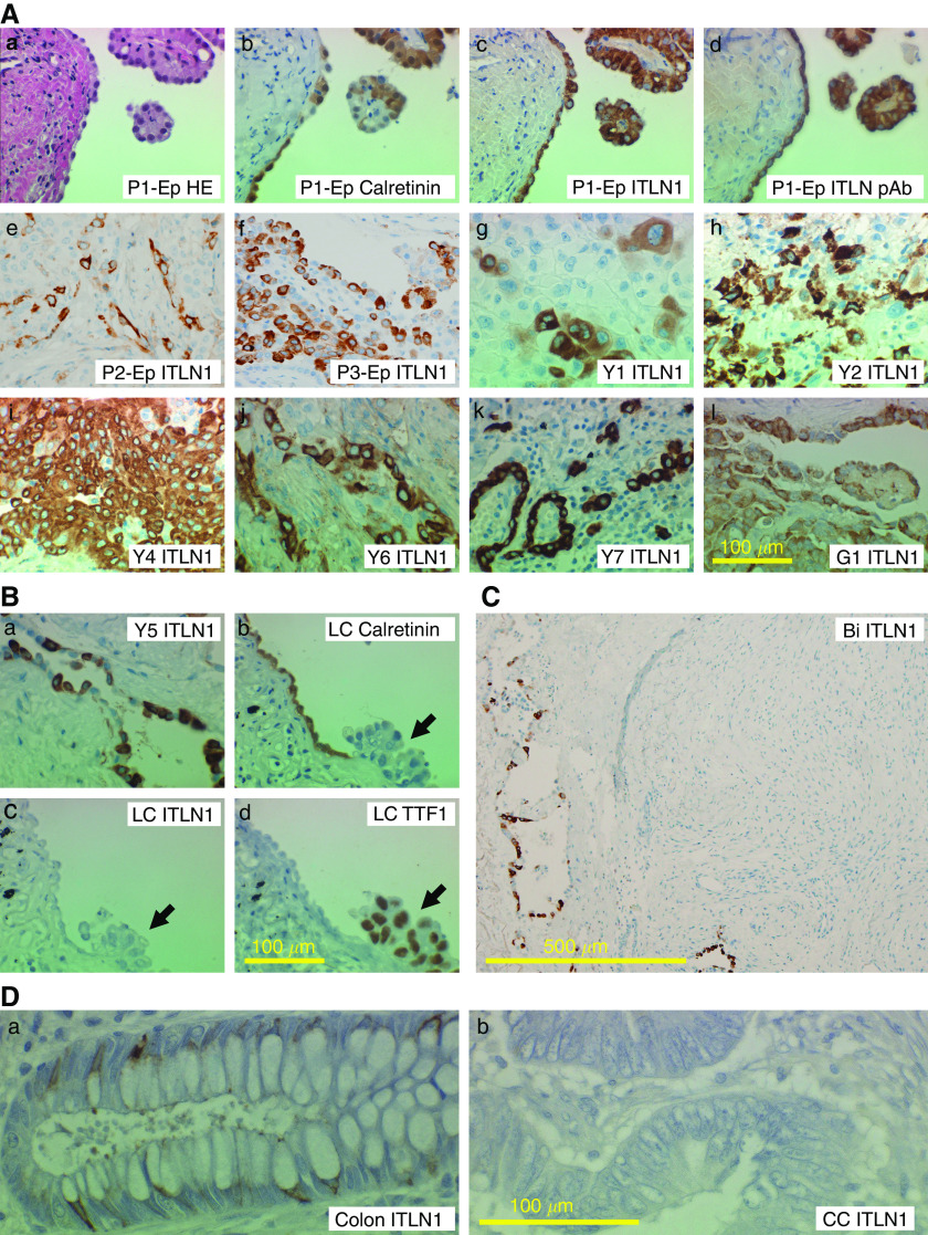
Figure 3.
Immunohistochemistry of intelectin-1 in MPM. Specimens of pleural biopsy were analysed by immunohistochemistry. Patients Y1, Y2, Y4, Y5, Y6, Y7, and G1 are identical to the ones in Table 1, respectively. Photographs were taken with a × 10 (panel C) or × 40 (others) objective lens. A scale bar is shown in the bottom side of each panel, representatively. (Aa) Haematoxylin–eosin (HE) staining of a specimen of an epithelioid-type MPM patient (P1-Ep). (Ab) Calretinin staining of MPM in the same specimen. (Ac) Intelectin-1 (ITLN1) staining of MPM in the same specimen with 15 : 3G9. (Ad) ITLN1 staining of MPM in the same specimen with anti-intelectin pAb. (Ae–Al) ITLN1 staining of MPM in eight epithelioid-type MPM patients (P2-Ep, P3-Ep, Y1, Y2, Y4, Y6, Y7, or G1) with 15 : 3G9 (Ae–Ak) or anti-intelectin pAb (Al). (Ba) ITLN1 staining of pleural surface MPM in an epithelioid-type MPM patient (Y5). (Bb) Calretinin staining of reactive mesothelial cells on a pleura invaded by lung adenocarcinoma in a lung cancer patient (LC). (Bc) No staining of the pleural cells in the same specimen with 15:3G9. (Bd) TTF-1 staining of pleura-invading lung adenocarcinoma in the same specimen. The arrows indicate the lung adenocarcinomas invading the pleura. (C) ITLN1 staining in a biphasic-type MPM patient (Bi). ITLN1 was stained with 15 : 3G9 in epithelioid-like MPMs in the left side, but not sarcomatoid-like MPMs in the centre and right side. (Da) ITLN1 staining of normal colonic goblet cells. (Db) No staining of colon adenocarcinoma in the identical patient (CC) with anti-intelectin pAb.

