Fig. 4.
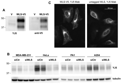
Localization of WLS to the ER. (A) Western blot using monoclonal antibody YJ5 recognizes transiently expressed WLS. WLS-V5-His was expressed in HEK293 cells and lysates were probed with mAb YJ5 or anti-V5 as indicated. V, lysates from cells transfected with vector alone. (B) WLS was expressed in multiple cancer cell lines. Whole-cell lysates from indicated cell lines (25 μg) transfected with control (SiCtr) or specific siRNA directed against WLS were probed with YJ5 Mab. (C) WLS was present in the ER. HeLa cells that transiently expressed C-terminally V5-His tagged or untagged WLS were stained with YJ5. Strong perinuclear staining, indicative of ER localization, was seen when untagged WLS was expressed. YJ5 immunoreactivity was seen very weakly in non-transfected cells (data not shown).

