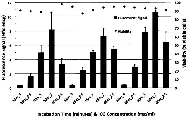
Figure 2.
Cell viability of human embryonic stem cells (hESCs) maintained postlabeling with indocyanine green (ICG). Fluorescent signal (units in efficiency) seen with optical imaging (OI) and viability (% viable cells) of hESCs, labeled with different ICG concentrations (0 = control as well as 0.5, 1.0, 1.5, 2.0, and 2.5 mg ICG/ml) for different incubation intervals (30, 45, and 60 min). Data are displayed as means and SD of triplicate samples containing 300,000 cells in a pellet per sample in 1 ml of KSR.

