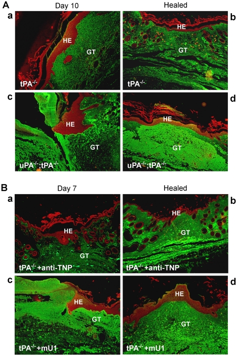
Figure 3. Cytokeratin and fibrin immunofluorescence stainings of wound areas from mU1- or control mAb (i.e. anti-TNP)-treated mice.
Double immunofluorescence staining of cytokeratin (546 nm, red) and fibrin/fibrinogen (488 nm, green) in wound sections. A, Wounds from tPA-deficient and uPA;tPA double-deficient mice during healing (left panel, 10 days post wounding) and after re-epithelialization (right panel). B, Micrographs of wounds isolated from control mAb-treated (upper panels) and mU1-treated (lower panels) mice during healing (left panel, 7 days post wounding) and upon healing (right panel, 21 days post wounding).HE; hyperproliferative epidermis, GT; granulation tissue.

