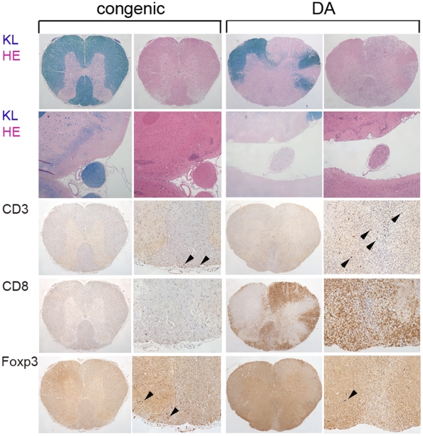
Figure 4. CNS histopathology demonstrates less inflammation and demyelination in DA.PVG-Eae23 than DA.
Spinal cord and brain sections of DA.PVG-Eae23 congenic rats (the two left columns) and DA rats (the two right columns) with the same MAX score. Sections were stained in the following order: spinal cord cross-sections (top row) and brain sections including optic nerve (row 2) were stained with Klüver (KL) and Hämalaun-Eosin (HE), respectively. Congenic rats revealed no signs of CNS inflammation and demyelination while DA rats showed severe loss of myelin and cell infiltration at the sight of the lesion in the spinal cord white matter, as well as at the optic nerve. Staining of the spinal cord sections against CD3 (row 3), CD8 (row 4) and Foxp3 (bottom row) revealed the higher recruitment of CD3+ T cells and CD8+ macrophages respectively to inflammatory lesions in the DA rats and higher recruitment of Foxp3+ T cells in DA.PVG-Eae23 congenic rats. Arrow heads point to cells with positive staining and indicate the relative number of positive cells present.

