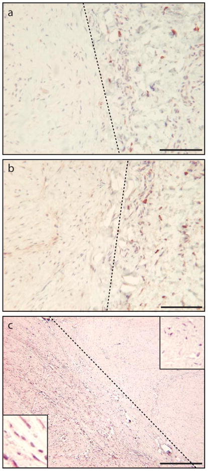
Figure 1. Distribution of SFRP1, SFRP2, and IGFBP5 immunostaining in keloid tissue.
(a) Active fibroblasts (left of dotted line) show no SFRP1 staining; inactive fibroblasts (right of dotted line) show positive staining; (b) Active fibroblasts (left of dotted line) show minimal SFRP2 staining; inactive fibroblasts (right of dotted line) show positive staining. Only keloid tissue left of dotted lines in a and b stains for type 1 procollagen, a marker of activated fibroblasts (data not shown); (c) Robust IGFBP5 immunoreactivity is seen only in area left of dotted line, which contains numerous elongated fibroblasts that appear to be actively growing and migrating. Weakly stained fibroblasts right of dotted line exhibit a rounded more mature phenotype and are sparsely distributed in matrix. Insets on lower left and upper right show high magnification field of each area. Scale bars for (a) and (b) = 100μm. Scale bar for (c) = 500μm.

