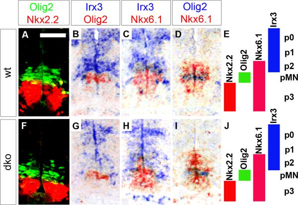
Fig. 2. VZ patterning in embryonic SC from wildtype mice and mice with SoxC deficiencies.
Immunohistochemistry with antibodies specific for Nkx2.2 and Olig2 (A,F) and in situ hybridizations with antisense riboprobes for Olig2, Nkx6.1, and Irx3 (B–D,G–I) were performed on transverse thoracic level sections of wildtype (wt) (A–D) and Sox4Δ/Δ Sox11lacZ/lacZ (dko) (F–I) embryos at 11.5 dpc to determine the integrity of the ventral VZ domains as summarized in E and J. In situ hybridization signals obtained with different probes on immediately adjacent sections were superimposed using Adobe Photoshop with the color of one of the signals being converted to red. A magnification of the signal displaying part of the VZ is shown. Scale bar in A is valid for all panels and corresponds to 50 μm.

