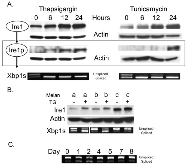
Figure 1.
(A) WT melanocytes were treated with 150nM thapsigargin or 10μg/mL tunicamycin for the indicated times, (B) mutant cells were treated with 150nM thapsigargin for 24 hours and (C), and WT melanocytes were treated with 15nM thapsigargin over an eight day period. Cells were lysed, proteins and RNA harvested and samples normalized for protein/RNA concentration prior to analysis. Ire1 expression and phosphorylation was monitored by Western blot analysis and Xbp1 splicing by RT-PCR as previously reported (Lin et al., 2007). Actin is shown as a loading control.

