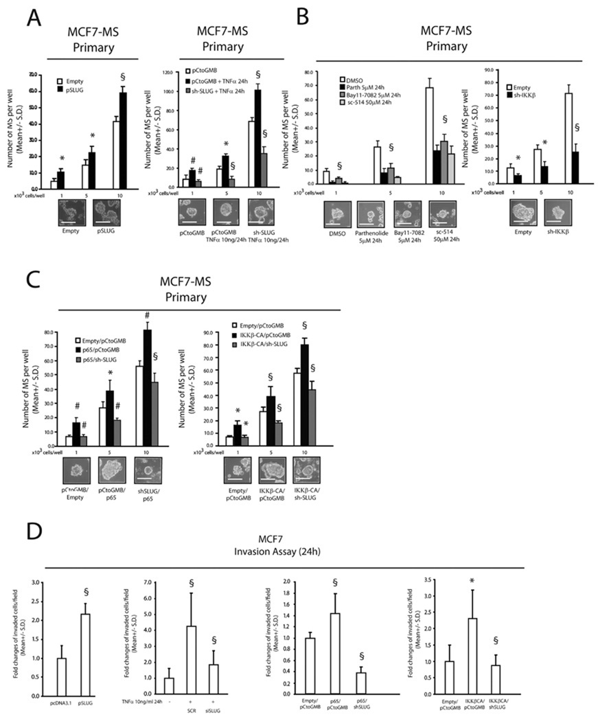
FIGURE 2. TNFα/NF-κB signalling promotes MS formation and invasive capacity of breast cancer cells via SLUG up-regulation.
(A): MS forming capacity of empty/pSLUG transiently transfected (1µg, 24h) MCF7 cells and pCtoGMB/ shSLUG stably transduced MCF7 cells exposed or less to TNFα (10 ng/ml, 24h); (B): MS forming capacity of MCF7 cells exposed to the IκBα degradation inhibitors Parthenolide or Bay 11-7082 (5µM, 24h each), the specific IKKβ inhibitor sc-514 (5µM, 24h each), or stably transduced with an IKKβ specific shRNA/empty expressing retroviral vector; (C): MS forming capacity of pCtoGMB/shSLUG MCF7 cells transduced with empty or p65/IKKβ-CA encoding vector, representative MS pictures are also reported. The scale bar inset corresponds to 100µm; (D): Invasion assay in pSLUG transfected (1µg), TNFα exposed (10ng/ml, 24h), p65/IKKβ-CA transduced pCtoGMB/shSLUG MCF7 cells. Data are presented as mean +/− S.D. of three replicates, p values of unpaired t tests: *<0.05, #<0.01 and § <0.005. The scale bar represents 100 µm.

