
Fig. 4. GIT1 functions downstream of RANK signaling.
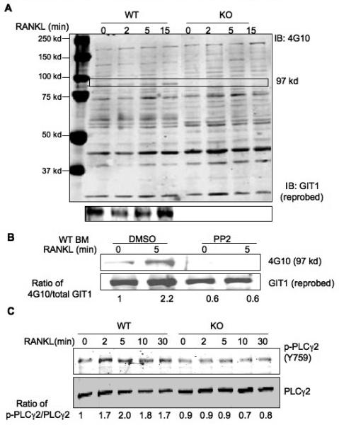
(A) GIT1 WT and KO BM cells were differentiated into OC by treatment with RANKL (50 ng/ml) and MCSF (20 ng/ml) for 7 days. Cells were serum starved for 6 hrs, stimulated with 100 ng/ml RANK for the indicated times. Cell lysates were harvested and probed with 4G10 phosphotyrosine antibody to visualize GIT1 phosphorylation (97 kd, arrow). Blots were then probed for GIT1 expression. (B) WT BM OC were treated with Src inhibitor PP2 (10 μM for 1 hr), stimulated with 100 ng/ml RANKL for 5 mins. GIT1 phosphorylation was probed using 4G10 antibody. Blots were reprobed for GIT1 expression. (C) GIT1 WT and KO OC on day 7 were starved and treated with RANKL (100 ng/ml) for indicated times. Lysates were immunoblotted with phoshorylated specific PLCγ2 (Y759) antibody (p-PLCγ2 Y759). Fold induction of normalized, phosphorylated protein vs time 0 of the control are shown.