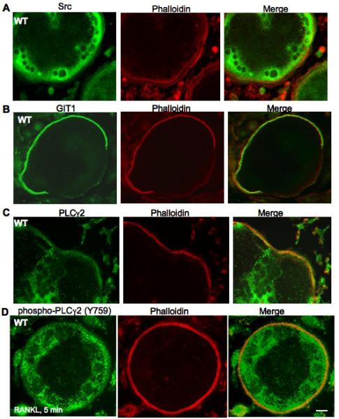
Fig. 5. Src, GIT1 and PLCγ2 localizes in the podosome belts.

WT KO BM cells were differentiated into OC by treatment with MCSF (20 ng/ml) and RANKL (50 ng/ml) for 7 days, fixed and stained for (A) Src, (B) GIT1 and (C) PLCγ2. (D) WT OCs were serum starved for 4 hrs and stimulated with RANKL (100ng/ml) for 5 mins, fixed and stained for phosphorylated form of PLCγ2 using phospho- PLCγ2 (Y759) antibody. Rhodamine-phalloidin was used to visualize actin rings. (Scale bar: 15 μm)
