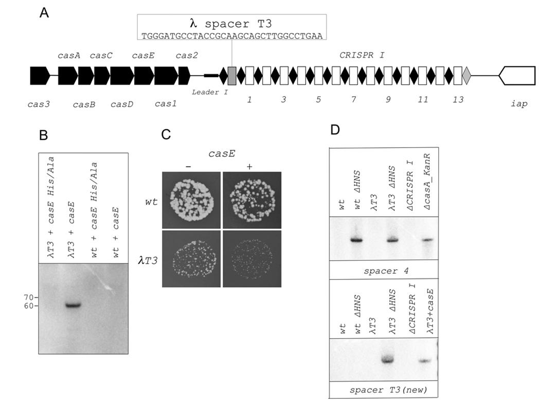
Figure 6. Function of genomic E. coli CRISPR in phage protection.
A. The structure of engineered CRISPR cassette in λT3 (BW39651) strain is shown (see Fig. 1A for details).
B. Northern blot analysis of processed transcripts using spacer T3-specific probe in RNA prepared from wild-type or λT3 strains carrying indicated casE expression plasmids.
C. The size or plaques formed by bacteriophage λ on indicated cell lawns. Identical dilutions of phage λ were deposited on lawns of indicated cells. Results of overnight growth at 30 °C are presented. The colors of plate photographs were inverted to make phage plaques better visible. The plaques appear white, while cell lawns appear black.
D. Northern blot analysis of processed transcripts using spacer 4 (top) and spacer T3 (bottom) specific probes in RNA prepared from wild-type or λT3 strains with or without the hns gene. ΔCRISPRI and casA_KanR are controls (see Fig. 1 and Fig. 4 legends).
E. Plaque formation by bacteriophage λ on indicated cell lawns. See panel C legend for details.

