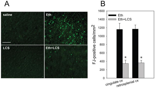
Fig. 4. The effects of LCS on ethanol-induced Fluoro-Jade C staining.
A: Mice treated with LCS/saline via ip were exposed to ethanol/saline as described in Materials and Methods. Twenty four hours after ethanol/saline exposure, mice were perfusion-fixed, and brain sections from four groups, saline only (saline), LCS only (LCS), ethanol only (Eth), and ethanol+LCS (Eth+LCS) group, were stained with Fluoro-Jade C. The images show the cingulate cortex area. The bar indicates 50 μm. B: Fluoro-Jade C (FJ) positive cells were counted in the cingulate cortex (cingulate CX) and in the retrosplenial cortex (retrosplenial CX) as described in Materials and Methods. *Significantly (p< 0.05) different between the ethanol (Eth) and ethanol+LCS (Eth+LCS) groups by Student’s t-test.

