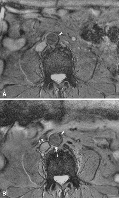
Figure 3.
Axial 2D gradient-echo MRI of a patient. Pre- (A) and postcontrast (B) images display the level of the aorta. On the precontrast image, the aortic wall is homogeneously hyperintense (A, arrowhead). Following SPIO administration, a pronounced signal loss of an area extending from the inner to the outer surface of the aortic wall can be seen (B, arrowhead). This vessel segment was considered positive. Note, however, that there is also a low-intensity ring at the interface between the aortic wall and lumen on the postcontrast image (B, long arrow). It is not possible to precisely determine whether the ring is truly confined to the aortic wall or to the lumen. This appearance is defined as a ring phenomenon, which is also seen in other vessels, such as the inferior vena cava (B, small arrowheads). Reproduced with permission from John Wiley & Sons67

