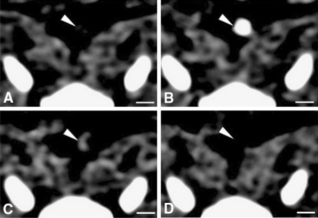
Figure 4.
Axial views of the same atherosclerotic plaque (white arrowheads) in the aorta of a rabbit, obtained by CT before (A), during (B) and 2 h after the injection of an iodine nanoparticle contrast agent (C) or a conventional iodine contrast agent (D). Adapted with permission from Macmillan Publishers Ltd49

