
FIGURE 2.
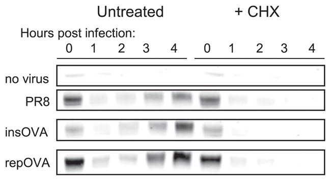
Functional NA protein synthesis begins 2–3 h p.i. DC2.4 cells were infected as in Fig. 1, cells were harvested at the indicated, times and total cell lysates were prepared for Western blot analyses using rabbit polyclonal Abs raised against the C terminus of the NA protein. Cells were either left untreated or incubated with CHX immediately p.i. The prominent band at ~50 kDa corresponding to the anticipated size of the NA monomers is shown.