
Figure 2.
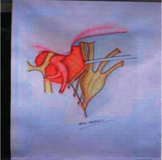
Drawing of the surgical anatomy. The intracavernous portion of the adenoma is fully exposed following lateral displacement of the released III cranial nerve using a nerve self-retaining retractor
Official websites use .gov
A
.gov website belongs to an official
government organization in the United States.
Secure .gov websites use HTTPS
A lock (
) or https:// means you've safely
connected to the .gov website. Share sensitive
information only on official, secure websites.
Drawing of the surgical anatomy. The intracavernous portion of the adenoma is fully exposed following lateral displacement of the released III cranial nerve using a nerve self-retaining retractor