
Figure 1.
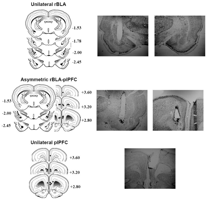
Schematic drawings representing coronal sections of the rBLA and plPFC subregions (left panels). Circles indicate the diameter (1.0 mm) of the theoretical diffusion of lidocaine from the cannulae tips of rats with unilateral rBLA (top), asymmetric rBLA/plPFC (middle), and unilateral plPFC (bottom) placements. All drawings are based on the atlas of Swanson (1992), with the anterior-posterior references measured from bregma. Each placement is shown at the midpoint of its anterior-posterior extent. Representative photomicrographs of asymmetric and unilateral cannulae tracks also are depicted (right panels).