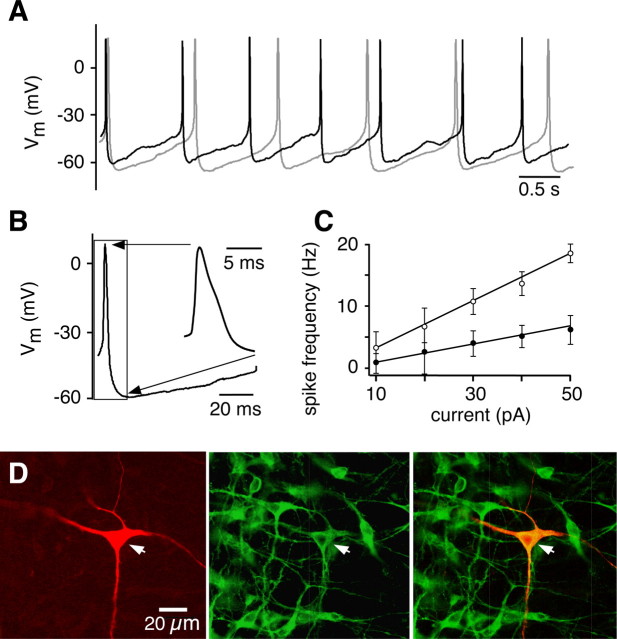
Figure 2.
Spontaneous firing of neurons in the n. raphé obscurus. A, Current-clamp recording from a representative raphé neuron firing at ∼1 Hz, in aCSF with [K+]o of 3 mm (gray trace) or 9 mm (black trace). B, Typical action potential shape, with visible shoulder and prominent AHP as shown in inset on the right, which is an expanded view of the region of the action potential bounded by the box on the left. C, Relationship between firing frequency and injected current for the first (○) and last (●) interspike intervals (n = 22 neurons) obtained during step injection of bias current. Difference in spike frequencies at a given current reflects spike frequency adaptation during sustained steps in bias current. D, Example of a regularly, tonically spiking raphé obscurus neuron filled with biocytin (1%) (Texas Red fluorescence, left). The same slice was stained for TpOH-immunoreactive neurons (Alexa Fluor 488 fluorescence, middle). Merged image (right) shows that the tonically spiking neuron was serotonergic.

