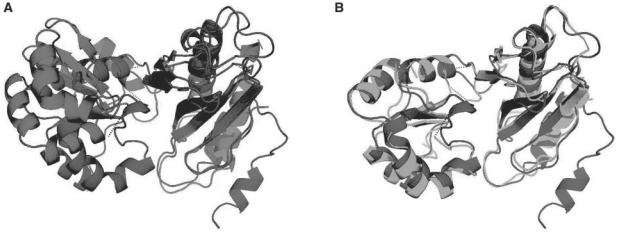
Fig. 4.
Comparisons between the ribosomal protein L1 mutant S179C [SCOP code d1ad2, in blue (black)] and the 50S ribosomal protein L1P from Methanococcus jannaschii [SCOP code d1cjsa, in green and red (gray)]. (A) The superposition of these two proteins according to the best rigid superposition possible for the matching found. It is apparent that only one domain from each protein is superimposed in this manner. Several local transfomations, however, allow a much better superposition (B).

