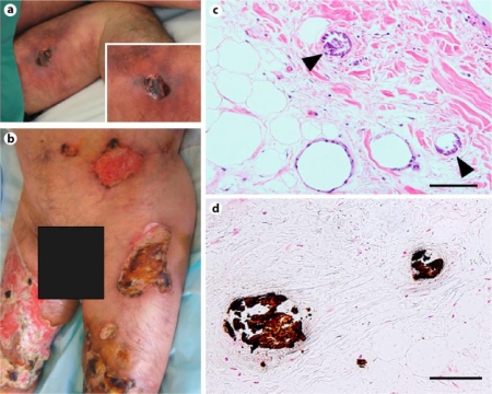
Fig. 1.
Calciphylaxis-induced skin changes at admission (a left thigh). A spread of the ulcer had expanded to the bilateral legs and trunk in a few months (b lower trunk and bilateral thighs). Histology of biopsy specimen from the ulcer on the left thigh showed classic findings of calciphylaxis including intimal hyperplasia with local fat necrosis (c) and calcium deposits (c, d arrowheads) in the media of small- and medium-sized arteries. c Hematoxylin-eosin stain; d von Kossa stain; scale bar = 100 μm.

