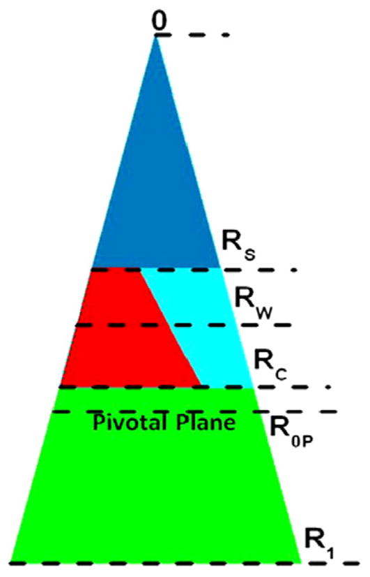
Fig. 10.

Schematic for the packing of lipid and water in the hexagonal II phase. The cylindrical water tubes extend in the direction perpendicular to the page. The center of each tube is at 0. The triangle extending from 0 to R1 represents the piece-of-pie space occupied by a monolayer of lipid and peptide and its associated water. The small dark blue (color online) triangle between 0 and RS (the steric radius) is occupied by water. The lower green trapezoid between R1 and RC (the Gibbs dividing surface for the hydrocarbon chains) represents the hydrophobic volume. The interfacial region between RS and RC represents the volume of the interfacial region; this is occupied by interfacial water (the right-hand light blue trapezoid) and the lipid headgroup and interfacial part of the peptide (the left-hand red trapezoid). The pivotal plane is at R0P. Radii are roughly to the scale of values for pure DOPE in Table 5.
