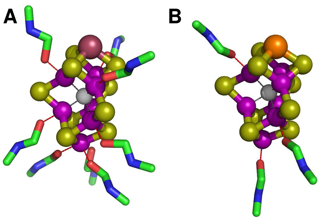
Figure 5.
Structural models of isolated FeVco (A) and FeMoco (B) in NMF. The structural models were adapted from the crystallographic coordinates of the MoFe protein2 but modified for distances on the basis of the EXAFS fits. The atoms are colored as follows: V, plum, Mo, orange; Fe, purple; S, lime; X (C, N or O), light gray; O, red; C, green; N, blue. The models reflect the EXAFS-derived coordination numbers of solvent molecules rather than the actual binding sites of these molecules.

